
Showing the measurement of length of manubrium of malleus 3. Length of... Download Scientific
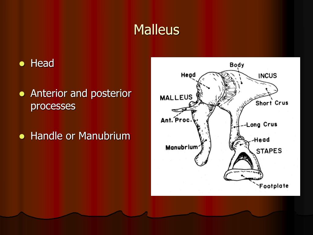
The malleus is divided into the head, neck, manubrium, anterior process, and lateral process. The incus is divided into the body, short crus, long crus, and lenticular process. The stapes is divided into the head (capitulum), neck, anterior crus, posterior crus, and footplate. [1]
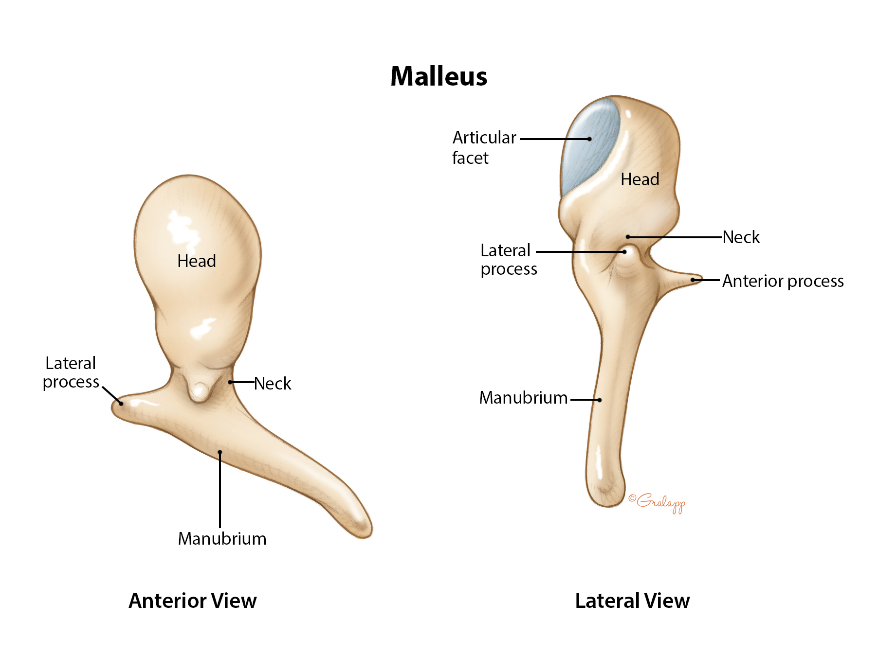
Middle Ear & Mastoid Oto Surgery Atlas
Background: The tympano-mallear connection (TMC) is the soft-tissue connection between the tympanic membrane (TM) and the manubrium of the malleus. Some studies suggest that its mechanical properties may have a substantial influence on the mechanics and transfer function of the middle ear.

Manubrium of right malleus of Vintana sertichi (UA 9972). A, in situ on... Download Scientific
This is the first of a two-part review that provides a practical approach to understanding temporal bone anatomy, localizing a pathologic process with a focus on inflammatory and neoplastic processes, identifying pertinent positives and negatives, and formulating a differential diagnosis. © RSNA, 2013 Learning Objectives:
:max_bytes(150000):strip_icc()/Malleus-1e2667df4ae74156a00144c2273fe1f4.jpg)
Malleus Anatomy, Function, and Treatment
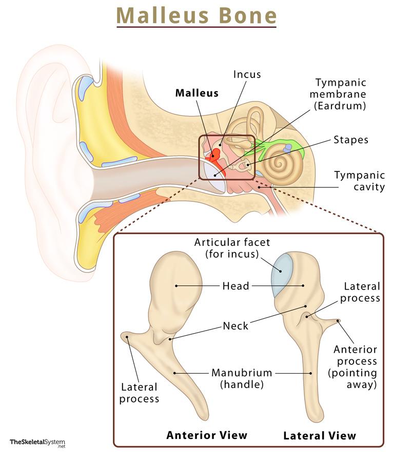
Malleus (ventral view) Malleus. The malleus, or hammer in Latin, develops from the first pharyngeal arch cartilage, like the mandible and maxilla jawbones. This small bone is connected with the tympanic membrane via its manubrium and with the incus via its articulating facet.The lateral process of the malleus is attached to the upper part of the tympanic membrane.

Malleus AnatomyZone
The manubrium mallei ( handle) is connected by its lateral margin with the tympanic membrane. It is directed downward, medialward, and backward; it decreases in size toward its free end, which is curved slightly forward, and flattened transversely.

تنه (دسته) استخوانچه چکشی اطلس الکترونیک آناتومی
The malleus (plural: mallei) is the most lateral middle ear ossicle, located between the tympanic membrane and the incus. Gross anatomy The malleus has a head, neck, and three distinct processes (manubrium (handle), anterior and lateral processes). The head is oval in shape, and articulates posteriorly with the incus by a small facet joint.
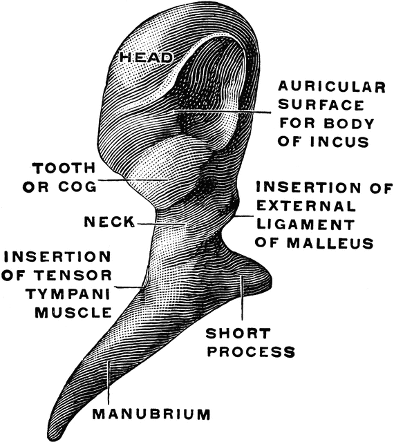
Malleus From Behind ClipArt ETC
The manubrium of the malleus is connected to the central region of the tympanic membrane and is responsible for the tympanic membrane's conical shape, as it pulls the tympanic membrane inward towards the middle ear cavity. From: Handbook of Clinical Neurophysiology, 2013 Add to Mendeley The Thorax Craig Cunningham,.

malleus incus stapes anatomy
Manubrium (Handle): This is the most notable landmark of the malleus, extending downwards as it curves and narrows towards its tip. The manubrium's lateral wall connects with the tympanic membrane. The small projection on its medial wall serves as the point of insertion for the tensor tympani muscle.
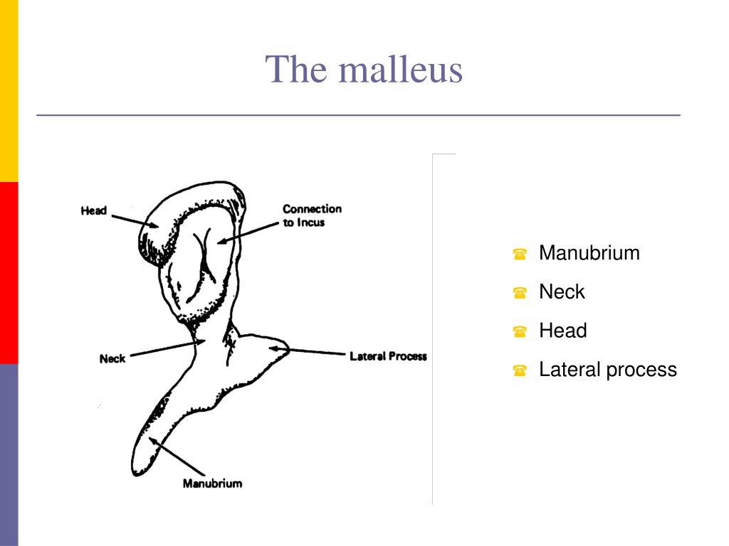
PPT CSD 3103 anatomy of speech and hearing mechanisms Hearing mechanisms Fall 2008 PowerPoint
The malleus is involved in only 2 per cent of traumatic lesions involving the middle ear. 2 Isolated fractures of the manubrium are even less common, and only 11 cases have been reported in the English language literature over the past 50 years (Table I). 1-5

The incus is positioned between the malleus and the stapes. In actual function, the bone serves
The malleus, or hammer, is a hammer-shaped small bone or ossicle of the middle ear. It connects with the incus, and is attached to the inner surface of the eardrum. The word is Latin for 'hammer' or 'mallet'. It transmits the sound vibrations from the eardrum to the incus (anvil). Structure The malleus is a bone situated in the middle ear.
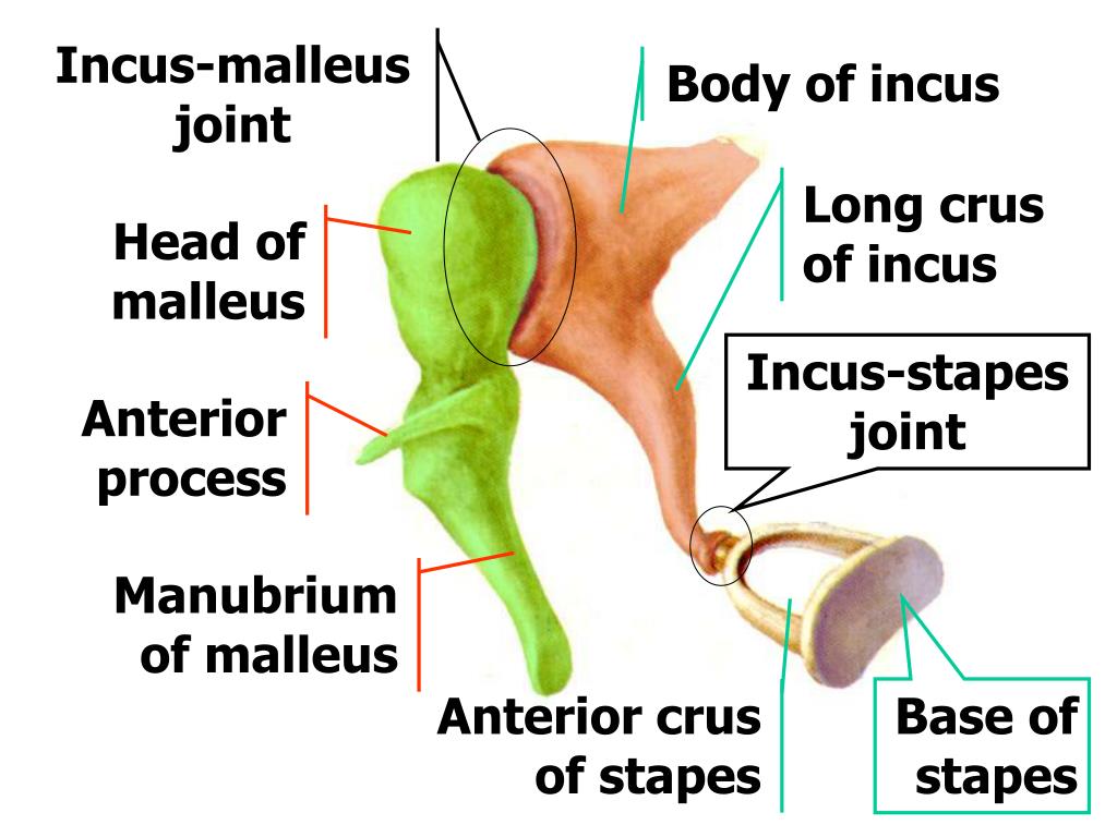
PPT Vestibulocochlear Organ (Ear) 前庭蜗器 ( 耳 ) PowerPoint Presentation ID6811281
The manubrium is a handle-like structure, as in the manubrium of the sternum or of the malleus. In Latin, it translates to "handle", which refers to that part of an object designed to hold. Let's take for instance the manubrium of the sternum, which is located in the upper part of the sternum (or breastbone).

—right malleus of neocnus dousman, uF 76364 A, medial view; B, lateral... Download Scientific
Background: There is no consensus why the manubrium of the malleus, as viewed clinically through the external ear canal, generally points downward and posteriorly. Objectives: To depict the alignment of the handle of the malleus, viewed clinically through the external auditory canal, relative to the zygomatic arch, the Frankfort plane, and a visual plane proxy and relative to the horizontal.
PPT Middle ear PowerPoint Presentation, free download ID3792146
It is widely accepted by developmental biologists that the malleus and incus of the mammalian middle ear are first pharyngeal arch derivatives, a contention based originally on classical embryology that has now been backed up by molecular evidence from rodent models.

Pin on Pathophysiology
The manubrium of the malleus is attached to the medial surface of the tympanic membrane, and it pulls its anterior and inferior portion medially, giving it a conical shape. The central point of maximum depression is called the umbo, and it marks the end of the manubrium. When observing the lateral surface of the tympanic membrane, superiorly.
Malleus Definition, Anatomy, & Functions With Diagram
Whyte et al. claimed that the manubrium of the malleus and the long process of the incus are separate from the bodies of these ossicles in the cartilaginous embryonic stages, the processes fusing with the bodies in human embryos of around 27-30 mm CRL. In the present study, with the single exception of H983, discussed below, the manubrium was.

a Section of the manubrium of malleus. b CT corresponding to a. 1... Download Scientific Diagram
A short manubrium of the malleus, demarcated by a distinctive bend from the base of the malleus, is known for docodontans ( Ji et al., 2006; Meng et al., 2015 ).